Zedu Weekly Wrap – 16 February 2024
It has been a romantic week…

but who needs roses when you have gel and the latest ultrasound system at hand 🙂
Highlights from the #FOAMed world to take a look at this week include:
- Michael Prats (@PratsEM) and the US GEL Podcast of SCUF interviews Part 2 (13 February).
- more from the team at MetroHealth and @mtabbut on Morel-Lavallee lesions (16 February)
Out the door now for the weekend…
Mike and Suean

16 February
Check out the latest video by@mtabbut
about using POCUS to evaluate for Morel-Lavallée Lesions. https://t.co/5N4MAfnhKf #POCUS #Ultrasound #MSKUltrasound #Orthopedics #orthotwitter #emergencymedicine #medtwitter #metroEUS pic.twitter.com/Ra9HEgBTfy— MetroHealth Emergency Ultrasound (@MH_EMultrasound) February 15, 2024
15 February
A week has gone past already! Yikes…
Today’s article looks at the utility of #POCUS from an anaesthetists point of view.
Check it out https://t.co/kIlDmkxX3k#ultrasound #ultrasoundtraining pic.twitter.com/m852FmiPWt
— Zedu Ultrasound Training @zedunow@med-mastodon.com (@zedunow) February 15, 2024
Ultrasound guided blocks for rib fractures in ED?
Look no further than @POCUSUK
Check out our cases with recent addition of original illustrations by @EveryOneNoOne👇🏽
Erector Spinae Plane Blockhttps://t.co/ew3nIFTebf
Serratus Anterior Plane Blockhttps://t.co/J1rfmzXLEp pic.twitter.com/nK5EruRxBB
— POCUS UK (@POCUSUK) February 15, 2024
Transoral and Transesophageal Point-of-Care Ultrasound in the Diagnosis of Bilateral Carotid Artery and Aortic Dissections
Neurocritical Care
DOI
10.1007/s12028-023-01909-9Sharelink: https://t.co/TCVyu0OFIc#POCUS #dissections pic.twitter.com/i4swqNBR4r
— Issac Cheong (@Issac80) February 14, 2024
@PLOSOne
14 February
Pediatric Pulmonology @PedPulmonol

Echocardiography Journal
Echocardiographic assessment of pediatric heart transplantation: A single-center experience in China

@TamingtheSRU
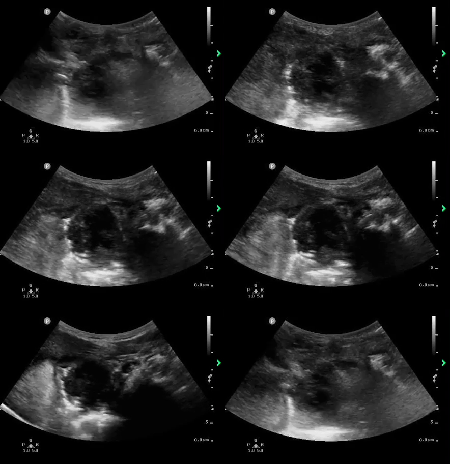
Ultrasound of the Month: Peritonsillar Abscess
Radiology Case Reports

Insights into Imaging @myESR

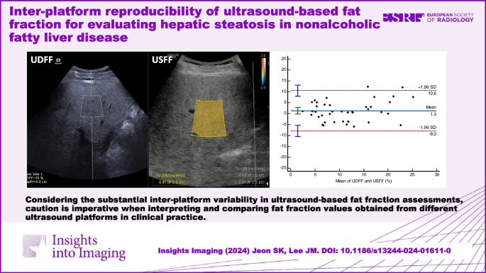
Insights into Imaging @myESR
13 February
US GEL PODCAST #155!
People/Progress/POCUS – #SCUF Interviews, Part 2 #POCUS #USGEL #FOAMed https://t.co/TZ7b13vAGt pic.twitter.com/oxmebM40kn
— Michael I. Prats (@PratsEM) February 12, 2024
@WFUMB

Radiology Case Reports
Ultrasound diagnosis of bowler’s thumb in a 60-year-old man

Clinical Practice and Cases in Emergency Medicine
@JCM_MDPI
@WINFOCUS

Make 2024 your year of ultrasound and check out our range of
scheduled and one-on-one options that will get you scanning like a pro.
12 February
Journal of Medical Radiation Sciences

@WINFOCUS
@WINFOCUS
Assessment of quadriceps muscle mass by ultrasound in the postoperative period of cardiac surgery

@SAEMonline
SAEM Clinical Images Series: One Month of Vaginal Bleeding

11 February
@ACEPNow
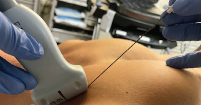
How To Perform an Ultrasound-Guided Clavipectoral Block
10 February
#FOAMed Case: Man With Abdominal Painhttps://t.co/hS5kebLoMo
•21yo man presents with acute onset r-lower quad pain with non-bloody emesis that started that morning;
•Rebound tenderness, guarding over McBurney’s point, positive psos sign and elevated WBC count. pic.twitter.com/4B48KgPqzW— ACEP Members (@ACEPNow) February 9, 2024
#POCUS
Are you using Resuscitative #TEE 🫀?
What views are needed?
How is it useful for #ACLS #CPR ?
Check out @ASRA_Society #ASRAPOCUS Spotlight article on #TEE #TOE !
Thx 🙏 @pperezde @JMorenoGarijo @UofTanesthesia 🙏
❤️ https://t.co/N9pGkadcrY @SCCM @ACEPNow @scahq @FTeranMD pic.twitter.com/bd7KsSKwXD— Hari (హరి) 🩺 Kalagara 🇮🇳 (@KalagaraHari) February 9, 2024
@WFUMB
Mitral Valve Segmentation and Tracking from Transthoracic Echocardiography Using Deep Learning

Insights into Imaging @myESR
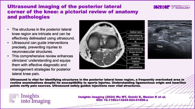
@JCM_MDPI
Multimodality Imaging in Right Heart Tumors: Proposed Algorithm towards an Appropriate Diagnosis
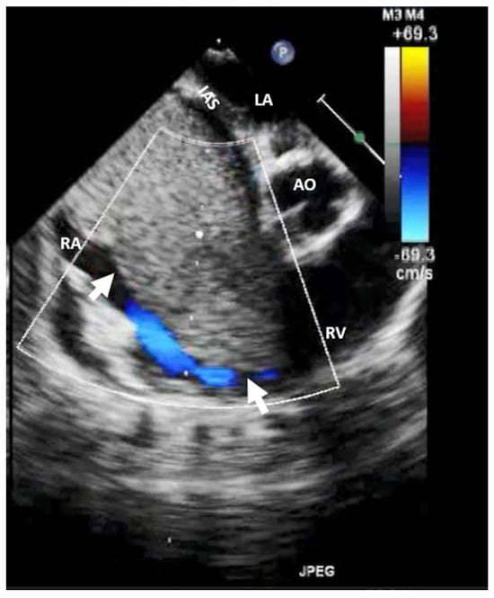
@JCM_MDPI
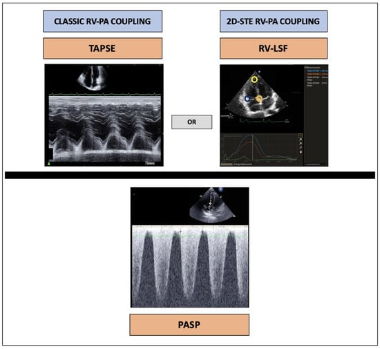
@MIT
An ultrasound sticker senses changing stiffness of deep internal organs
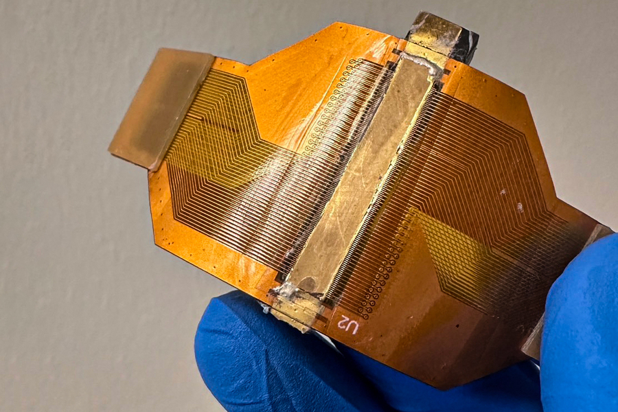
The Journal of Maternal-Fetal & Neonatal Medicine @tandfonline
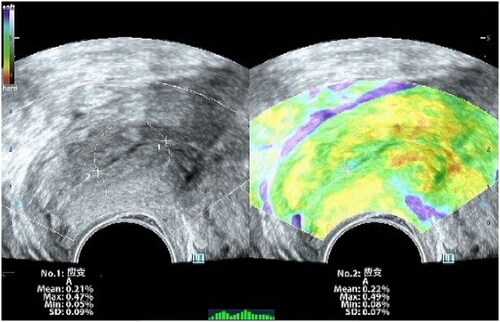
The Journal of Maternal-Fetal & Neonatal Medicine @tandfonline
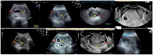
Ultrasound Int Open
Esophageal Leiomyoma: Case Report

You may also like
Zedu Weekly Wrap – 9 October 2026
Zedu Weekly Wrap – 2 October 2026