Zedu Weekly Wrap – 28 November 2025
December is around the corner…
 which means everyone we’ve been talking to have been turning their attention to matters of high priority – namely holidays and shopping. While we haven’t had the personal planning skills to make the most of Black Friday discounts, we have been busy putting our professional skills to good use to make sure everything is set and rady for 2026.
That being said – our calendar for the full year is ready and waiting for your review, with hundreds of days of ultrasound training open and available now. There are courses for every medical specialty and clinical application; and if there isn’t something that suits be sure to get in touch. We’re famous for our flexibility so reach out – we’ll do our best to get you scanning with confidence ASAP. So start planning and make 2026 your year of ultrasound.
Also be sure to add some reading to your weekend plans – highlight in the news this week 🚀✨
- Myocardial remodelling in aortic regurgitation: time to think beyond volumes and function? from the European Heart Journal (26 November)
- Left Ventricular Ejection Fraction Assessed by a General Practitioner Using AI-Guided Point-of-Care Ultrasonography: Feasibility and Diagnostic Agreement With Conventional Echocardiography from teh Journal of Diagnostic Medical Sonography (23 November)
Time to go plan something…
See you next week.
Mike and Suean
P.S. – our office will be closed over the period 19 December to 11 January.
28 November
Radiology Case Reports

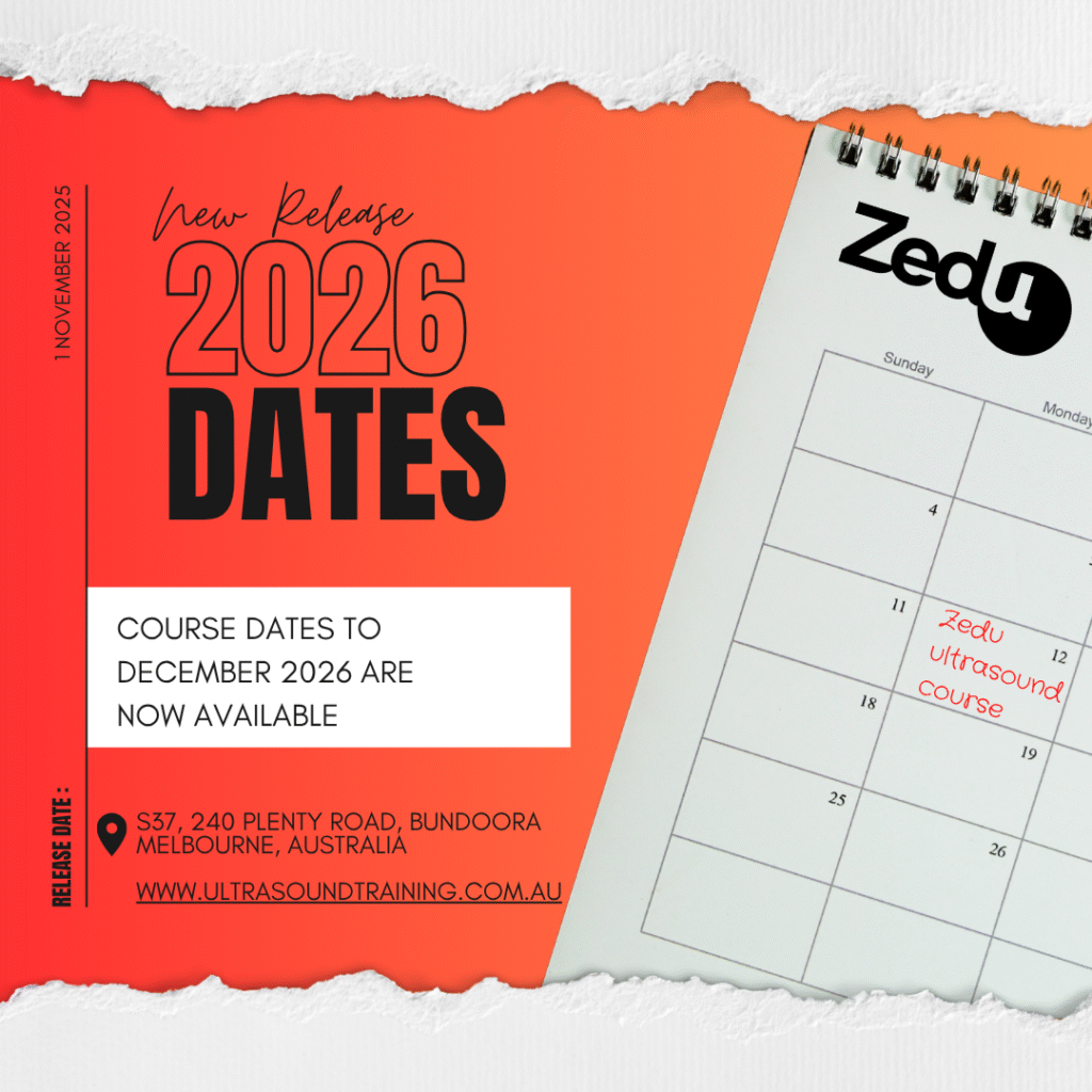
Make 2026 your year of ultrasound and check out our range of
scheduled and one-on-one options that will get you scanning like a pro.
Plus – as the cultural capital of Australia – why not tie in a bit of training with a touch of AO tennis, speed through at the Melbourne GP or have a laugh at the International Comedy Festival.
If you know the value of protected learning time, secure your place early.
P.S. – don’t forget – if something doesn’t quite fit your learning objectives or availability we can design and deliver a program that meets your needs. Don’t tell everyone though – availability is limited.

27 November
🚨#POCUS Abdo Education Review Studies show 🩸FAST/free fluid focus 🎉100% ⬆️ in competency 📚Need a blend of teaching methods 🛑Critical Gap- NO study compared methods head-2-head: we need efficiency data How do you get trainees from theory➡️bedside confidence? tinyurl.com/y2zet7r2 #ultrasoundtraining
— Zedu Ultrasound Training (@zedunow.bsky.social) 27 November 2025 at 09:53
Point‐Of‐Care Ultrasound in the Assessment of Cerebral Arteriovenous Malformations
Journal of Clinical Ultrasoundhttps://t.co/mfb7o88K7t
Shareable link: https://t.co/hmEyfLQf6d#POCUS #Neurosonology #TCD pic.twitter.com/tIhyfLmphb
— Issac Cheong (@Issac80) November 27, 2025
Radiology Case Reports
Role of ultrasound in the prenatal diagnosis of sacrococcygeal teratoma: A case study

Journal of Clinical Ultrasound

Journal of Clinical Ultrasound
A Comparative Study of Ultrasonography and CT Venography in the Diagnosis of Budd-Chiari Syndrome

26 November
Our FASTEMI study is now published! 🎉
This quick and practical POCUS protocol in STEMI can be truly decisive: it guides management, identifies critical differential diagnoses, and achieves a 97% negative predictive value for in-hospital mortality.https://t.co/OqRMxgCmwP pic.twitter.com/fPGANU8CPj— André B Amon (@AndreAmonMD) November 26, 2025
Applied Sciences
Recent Advances in B-Mode Ultrasound Simulators

European Heart Journal
Myocardial remodelling in aortic regurgitation: time to think beyond volumes and function?

25 November
There’s no missing Australia’s 🇦🇺 home of #ultrasound
Perfect weather for a perfect reveal
With all signs pointing to Melbourne the time to book in your 2026 #ultrasoundtraining is nowhttps://t.co/73GoKnT7g0 #POCUS #meded pic.twitter.com/83ioSdITRG— Zedu Ultrasound Training @zedunow@med-mastodon.com (@zedunow) November 25, 2025
The heart point sign: a bedside ultrasound marker of left-sided pneumothorax in critically ill patients−a case series
Journal of Ultrasound
DOI: 10.1007/s40477-025-01099-4
Shareable link: https://t.co/oJUTJfgXwp#POCUS #LUS #Pneumothorax pic.twitter.com/U5rsZhdung
— Issac Cheong (@Issac80) November 24, 2025
Radiology Case Reports

24 November
PLOSOne
23 November
Visual Journal of Emergency Medicine

The American Journal of Emergency Medicine

Journal of Diagnostic Medical Sonography

22 November
The Ultrasound Journal

AJOG
A critical review of placental function evaluation near term using Doppler ratios

Echocardiography Journal
Does Left Atrial Size and E/e′ Predict Outcomes in Hypertrophic Cardiomyopathy?

ISUOG

Journal of Clinical Medicine

Annals of Intensive Care
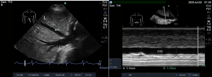
Ultrasound indicators of organ venous congestion: a narrative review
P.S. – don’t forget our brand-new headquarters are located at:
Suite 37 (Level 4), 240 Plenty Road, Bundoora Melbourne VIC 3083
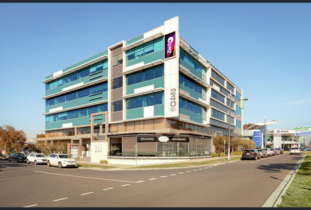
This isn’t just a change of address – it’s the start of something bigger.
 |
 |

You may also like

Zedu Weekly Wrap – 9 October 2026
Zedu Weekly Wrap – 2 October 2026